

Percutaneous Endoscopic Lumbar Discectomy (PELD): Bipolar Radiofrequency Hemostasis, Recovery, and O
Percutaneous Endoscopic Lumbar Discectomy (PELD) is a minimally invasive spine surgery technique used to treat lumbar disc herniation with smaller incisions, reduced tissue damage, and faster recovery compared with traditional open surgery.
A key technology supporting PELD is bipolar radiofrequency hemostasis, which enables precise bleeding control, tissue ablation, and annular sealing during endoscopic spine surgery.
What Is PELD?
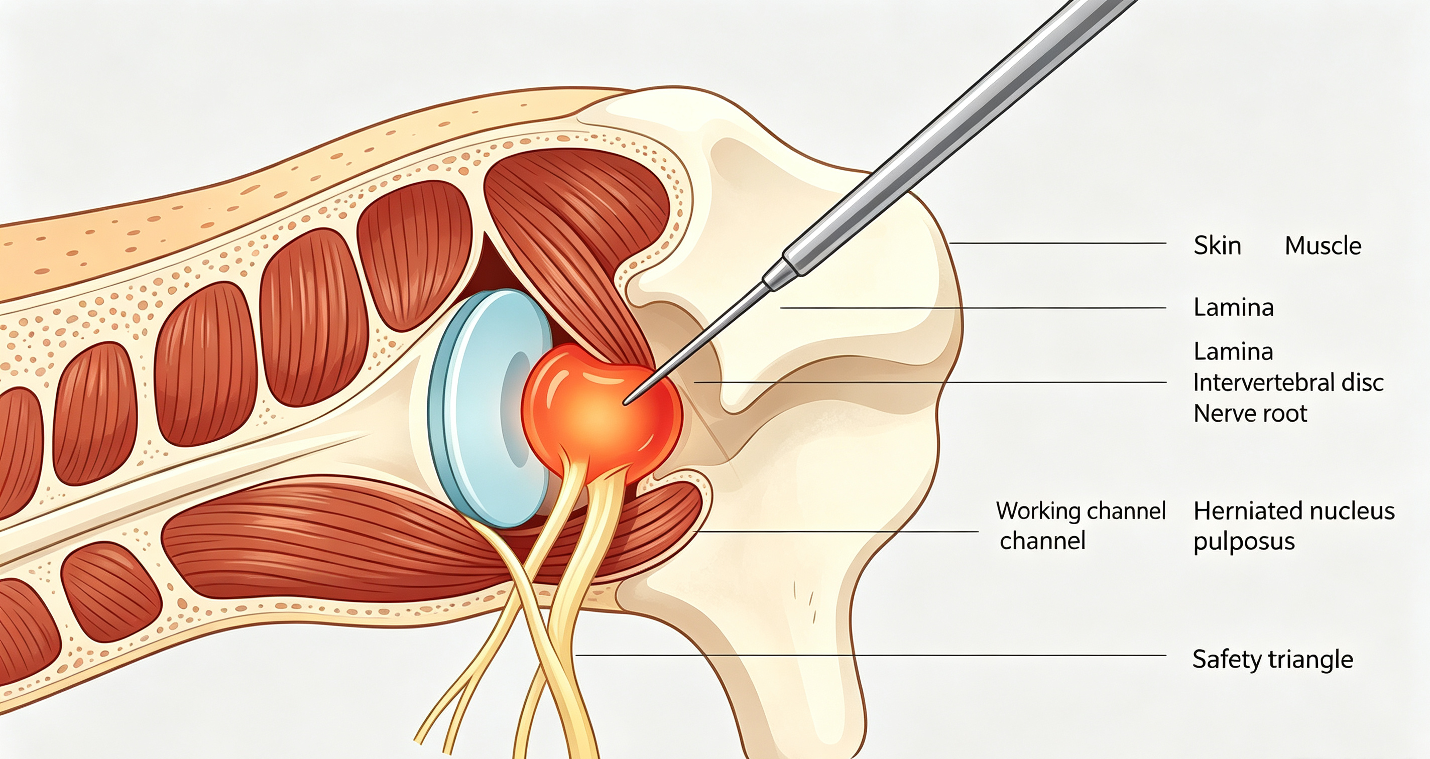
PELD removes herniated disc material through a small 7–10 mm incision using an endoscope and specialized instruments. Unlike conventional open discectomy, it avoids extensive muscle stripping and helps preserve normal spinal structures.
The procedure is usually performed under local anesthesia with sedation, allowing patients to communicate with the surgeon during surgery and improving safety during nerve decompression.
Indications for PELD
PELD is commonly used for patients with:
· Lumbar disc herniation causing radiculopathy
· Recurrent disc herniation after previous surgery
· Selected cases of spinal stenosis
· Symptoms that persist after conservative treatment
It is particularly suitable for patients seeking faster recovery or those who may have higher risks associated with general anesthesia.
Bipolar Radiofrequency Hemostasis in PELD
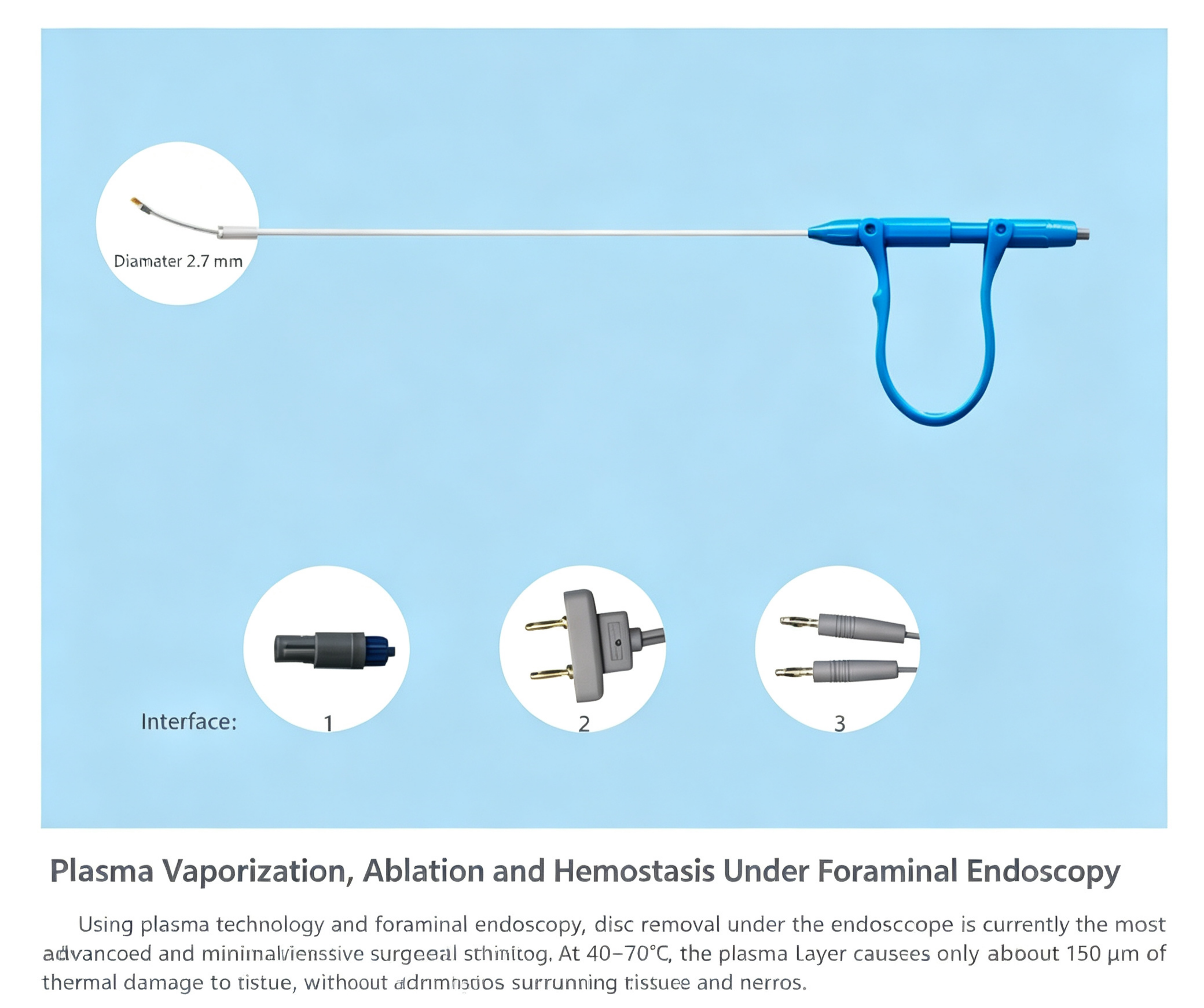
During PELD, continuous saline irrigation maintains a clear surgical field. The bipolar RF probe provides multiple functions:
· Coagulation of epidural and soft tissue bleeding
· Shrinkage of disc tissue
· Ablation of residual herniated tissue
· Removal of inflammatory and granulation tissue
· Annular sealing to help reduce recurrence risk
Compared with conventional electrosurgical techniques, bipolar radiofrequency delivers controlled energy at lower temperatures, minimizing thermal damage to surrounding nerves and tissues.
Advanced systems with bipolar turbo mode can provide enhanced hemostatic performance for challenging bleeding conditions.
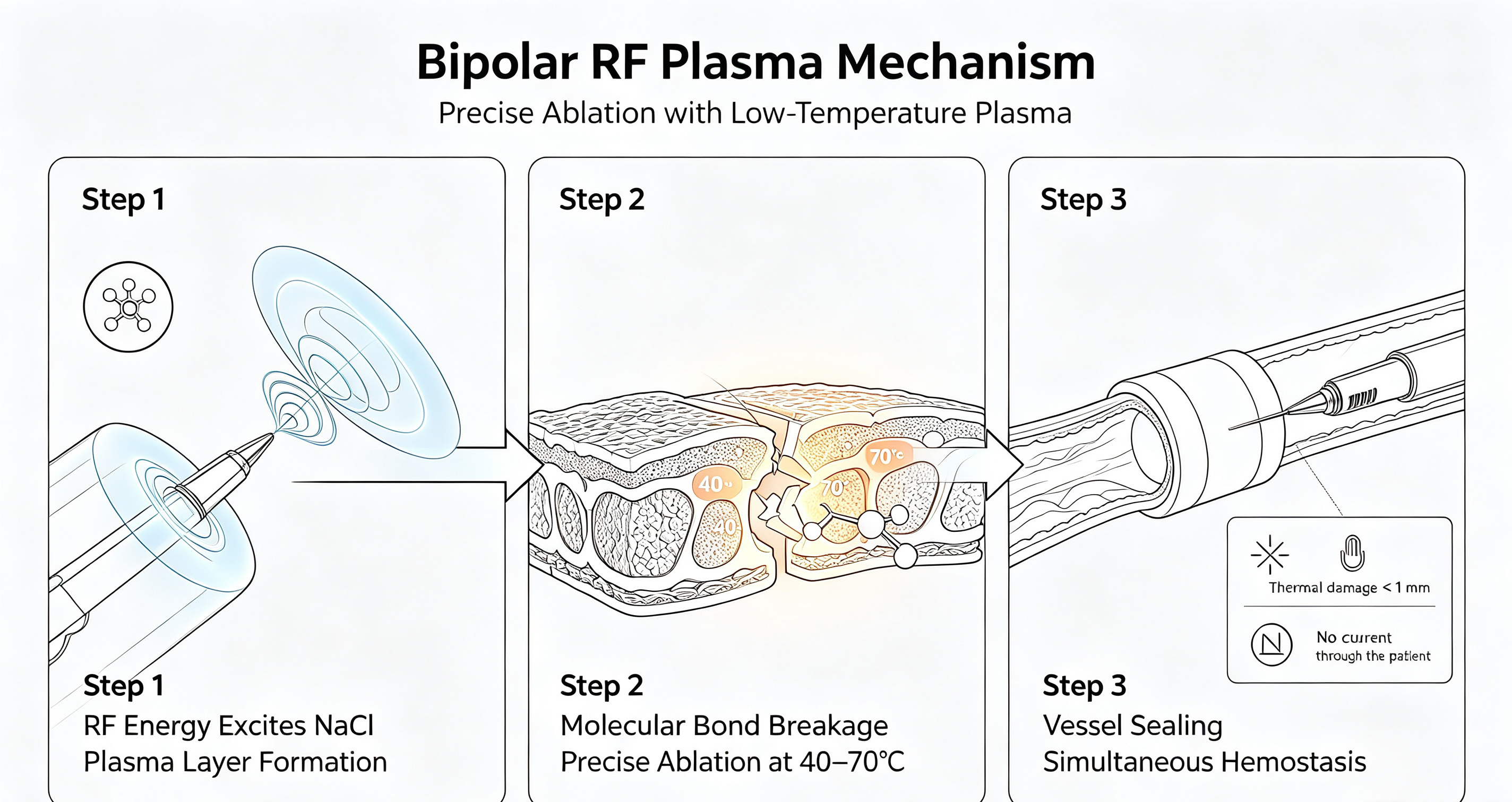
Importance of Hemostasis in Endoscopic Spine Surgery
In minimally invasive spine surgery, even small amounts of bleeding can affect visualization and surgical accuracy. Effective bipolar radiofrequency hemostasis helps maintain a clear operative field, improve efficiency, and enhance patient safety.
PELD vs. Open Surgery
Aspect | PELD | Open Surgery |
Incision | 7–10 mm | Several centimeters |
Anesthesia | Local + sedation | General anesthesia |
Muscle damage | Minimal | Extensive |
Hospital stay | 1–3 days | Several days or longer |
Recovery | 3–6 weeks | 3–6 months |
Spinal stability | Better preserved | Higher risk of disruption |
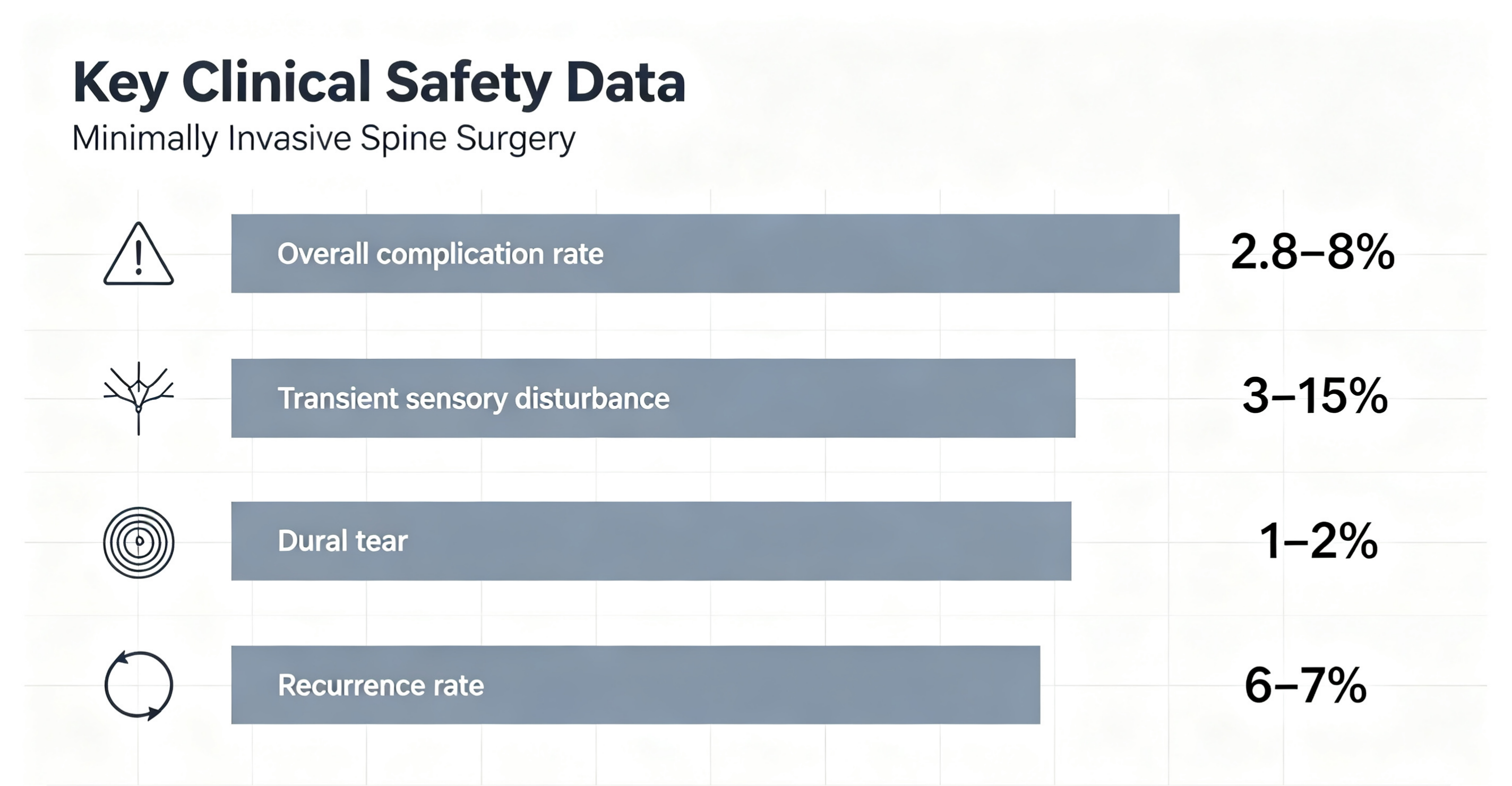
Clinical studies show that PELD provides comparable long-term outcomes to traditional surgery. Reported recurrence rates are generally around 6–7%, with complication rates approximately 2.8–8%.
PELD Procedure Overview
1. Imaging and Planning
MRI and CT scans are used to identify the affected disc level and determine the surgical approach.
2. Endoscopic Access
Under C-arm fluoroscopy guidance, the surgeon creates a working channel through a small incision.
3. Disc Removal
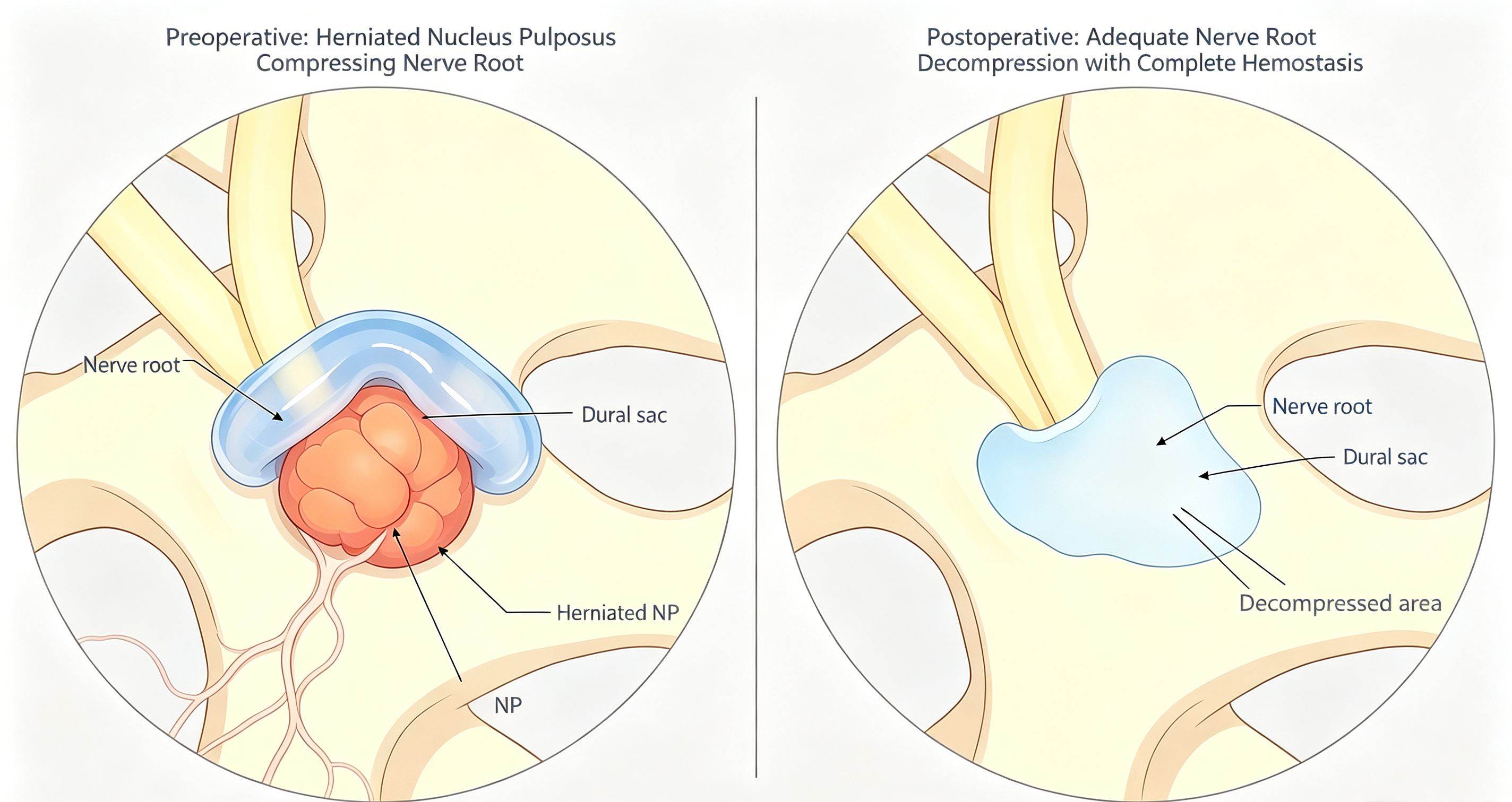
The herniated nucleus pulposus is removed under direct endoscopic visualization.
4. Bipolar Radiofrequency Hemostasis and Annuloplasty
The bipolar RF probe controls bleeding, removes residual tissue, and seals the annular defect to improve visualization and reduce recurrence risk.
5. Closure
The incision is closed with a small stitch or adhesive strip.
The procedure usually takes approximately 30–60 minutes.
Recovery After PELD
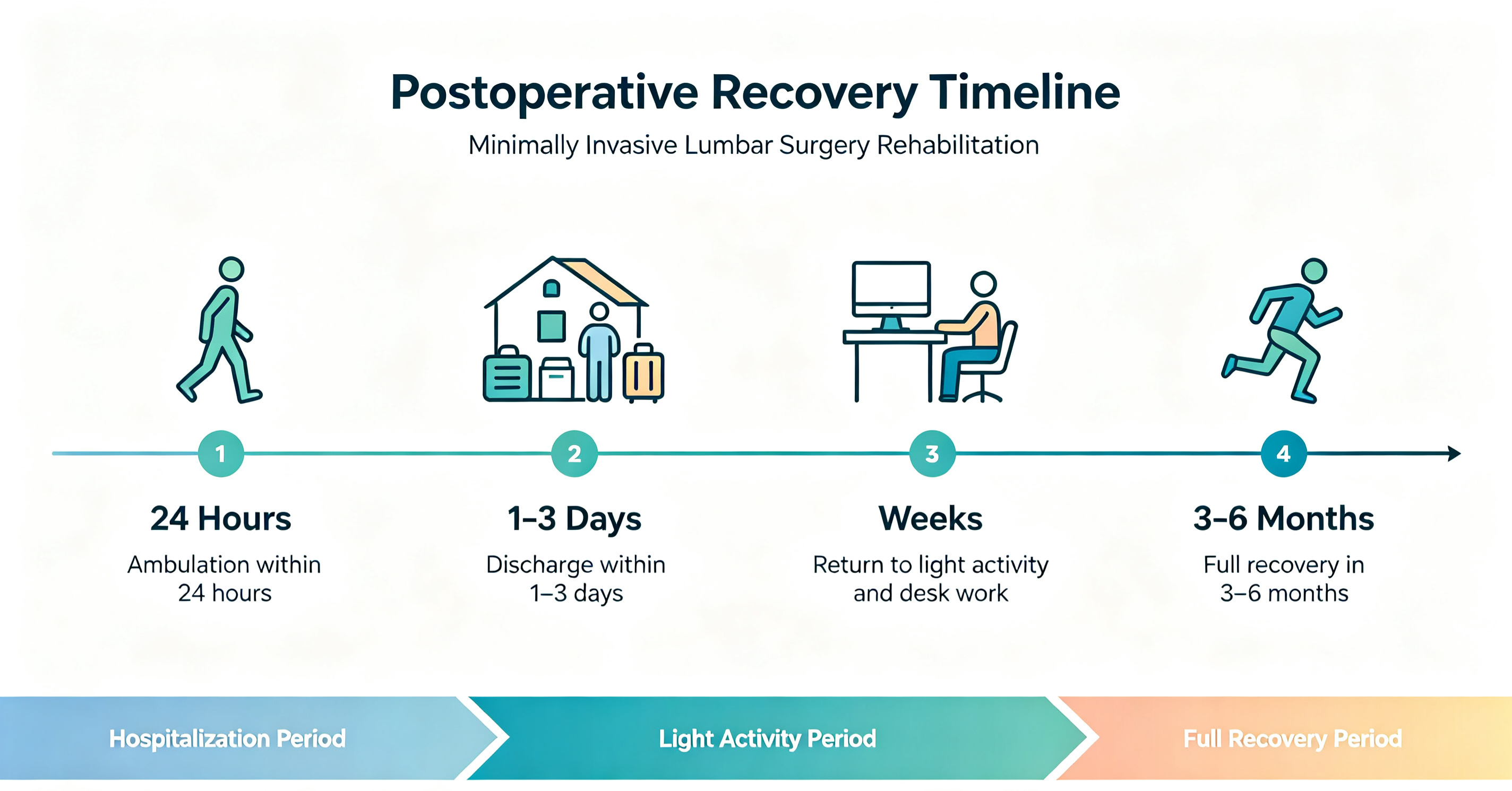
PELD allows a relatively rapid recovery:
· Patients can usually walk within 24 hours
· Hospital discharge typically occurs within 1–3 days
· Light activities may resume within several weeks
· Full recovery generally occurs within 3–6 months
Patients should avoid heavy lifting, excessive bending, and twisting during early recovery.
Risks and Complications
Although PELD is considered safe, possible complications include:
· Transient dysesthesia caused by nerve irritation (approximately 3–15%)
· Dural tear (about 1–2%)
· Recurrent disc herniation (around 6–7%)
· Rare infection or nerve injury
Most complications are temporary and manageable with proper treatment.
Conclusion
Percutaneous Endoscopic Lumbar Discectomy (PELD) with bipolar radiofrequency hemostasis represents an important advancement in minimally invasive spine surgery.
By combining a small incision, local anesthesia, precise endoscopic visualization, and advanced bipolar radiofrequency hemostasis technology, PELD provides effective treatment for lumbar disc herniation while reducing tissue damage and accelerating recovery.
For patients with persistent symptoms despite conservative treatment, PELD offers a valuable surgical option to discuss with a spine specialist.